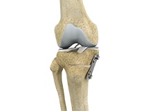
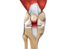
Knee Anatomy
The knee is a complex joint made up of different structures - bones, tendons, ligaments, and muscles. They all work together to maintain the knee’s normal function and provide stability to the knee during movement.
Having a well-functioning healthy knee is essential for our mobility and ability to participate in various activities. Understanding the anatomy of the knee enhances your ability to discuss and choose the right treatment procedure for knee problems with your doctor.
Bones of the Knee
The knee is a hinge joint made up of two bones, the thighbone (femur) and shinbone (tibia). There are two round knobs at the end of the femur called femoral condyles that articulate with the flat surface of the tibia called the tibial plateau. The tibial plateau on the inside of the leg is called the medial tibial plateau and on the outside of the leg, the lateral tibial plateau.
The two femoral condyles form a groove on the front (anterior) side of the knee called the patellofemoral groove. A small bone called the patella sits in this groove and forms the kneecap. It acts as a shield and protects the knee joint from direct trauma.
A fourth bone called the fibula is the other bone of the lower leg. This forms a small joint with the tibia. This joint has very little movement and is not considered a part of the main joint of the knee.
Articular Cartilage and Menisci of the Knee
Movement of the bones causes friction between the articulating surfaces. To reduce this friction, all articulating surfaces involved in the movement are covered with a white, shiny, slippery layer called articular cartilage. The articulating surface of the femoral condyles, tibial plateaus and the back of the patella are covered with this cartilage. The cartilage provides a smooth surface that facilitates easy movement.
To further reduce friction between the articulating surfaces of the bones, the knee joint is lined by a synovial membrane that produces a thick clear fluid called synovial fluid. This fluid lubricates and nourishes the cartilage and bones inside the joint capsule.
Within the knee joint, between the femur and tibia, are two C-shaped cartilaginous structures called menisci. Menisci function to provide stability to the knee by spreading the weight of the upper body across the whole surface of the tibial plateau. The menisci help in load-bearing i.e. it prevents the weight from concentrating onto a small area, which could damage the articular cartilage. The menisci also act as a cushion between the femur and tibia by absorbing the shock produced by activities such as walking, running and jumping.
Ligaments of the Knee
Ligaments are tough bands of tissue that connect one bone to another bone. The ligaments of the knee stabilize the knee joint. There are two important groups of ligaments that hold the bones of the knee joint together, collateral and cruciate ligaments.
Collateral ligaments are present on either side of the knee. They prevent the knee from moving too far during side to side motion. The collateral ligament on the inside is called the medial collateral ligament (MCL) and the collateral ligament on the outside is called the lateral collateral ligament (LCL).
Cruciate ligaments, present inside the knee joint, control the back-and-forth motion of the knee. The cruciate ligament in the front of the knee is called anterior cruciate ligament (ACL) and the cruciate ligament in the back of the knee is called posterior cruciate ligament (PCL).
Muscles of the Knee
There are two major muscles in the knee - the quadriceps and the hamstrings, which enable movement of the knee joint. The quadriceps muscles are located in front of the thigh. When the quadriceps muscles contract, the knee straightens. The hamstrings are located at the back of the thigh. When the hamstring muscles contract, the knee bends.
Tendons of the Knee
A tendon is a tissue that attaches a muscle to a bone. The quadriceps muscles of the knee meet just above the patella and attach to it through a tendon called the quadriceps tendon. The patella further attaches to the tibia through a tendon called the patella tendon. The quadriceps muscle, quadriceps tendon, and patellar tendon all work together to straighten the knee. Similarly, the hamstring muscles at the back of the leg are attached to the knee joint with the hamstring tendon.
-
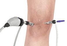
Knee Arthroscopy
Knee arthroscopy is a common surgical procedure performed using an arthroscope, a viewing instrument, to diagnose or treat a knee problem. It is a relatively safe procedure and you will usually be discharged from the hospital on the same day of surgery.
-
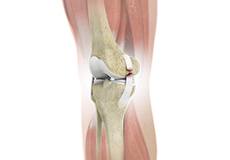
ACL Reconstruction
ACL (anterior cruciate ligament) reconstruction is a commonly performed surgical procedure. With recent advances in arthroscopic surgery, it can now be performed with minimal incision and low complication rates.
-
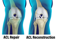
ACL Reconstruction Surgery: Graft Options
In the majority of patients who have torn their anterior cruciate ligament (ACL), surgery is recommended. The ACL has been shown to not heal on its own nor when it is repaired (surgically sutured or re-attached).
-
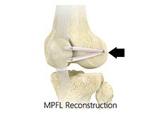
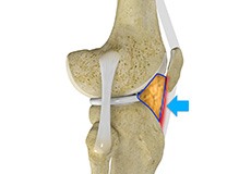
Medial Patellofemoral Ligament Reconstruction
The medial patellofemoral ligament can rupture or get damaged when there is patellar lateral dislocation. Dislocation can be caused by a direct blow to the knee, twisting injury to the lower leg, strong muscle contraction, or because of a congenital abnormality such as shallow or malformed joint surfaces.
-
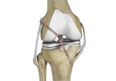
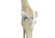
Multiligament Reconstruction of the Knee
Multiligament knee reconstruction is a surgical procedure to repair or replace two or more damaged ligaments of the knee joint. The surgery can be performed using minimally invasive techniques.
-
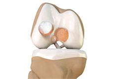
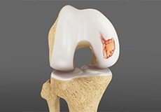
Cartilage Replacement
Cartilage replacement is a surgical procedure performed to replace the worn-out cartilage with new cartilage.
-
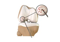
Cartilage Microfracture
Cartilage microfracture is a surgical procedure performed to replace the worn-out articular cartilage with new cartilage.
-
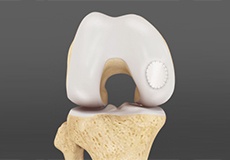
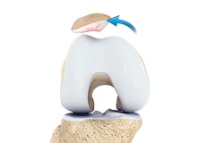
Knee Cartilage Restoration
Knee cartilage restoration is a surgical technique to repair damaged articular cartilage in the knee joint by stimulating new growth of cartilage or by transplanting cartilage into areas with defects in order to relieve pain and restore normal function to the knee.
-
Knee Osteotomy
Knee osteotomy is a surgical procedure in which the upper part of shinbone (tibia) or lower part of thighbone (femur) is cut and realigned. It is usually performed in arthritic conditions affecting only one side of your knee.
-
Meniscal Surgery
Meniscal surgery is a surgical procedure employed for the treatment of torn or damaged meniscal tissues in the knee. It is mostly performed as a minimally invasive keyhole procedure.
-
Meniscus Root Repair
Meniscus root repair is a surgery performed to repair a torn meniscus root. Meniscal repair may be performed either by open surgery under direct vision or minimally invasively using an arthroscope that can be inserted into the knee through a very small key-hole incision to locate and repair the damaged meniscus.
-
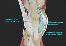
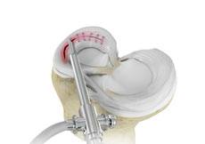
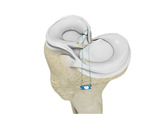
Patellofemoral Realignment
Patellofemoral realignment is a surgical procedure performed to treat symptomatic patellofemoral instability that does not respond to nonsurgical treatment measures.
-
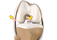
ACL Tears
The anterior cruciate ligament (ACL) is one of the major ligaments of the knee. It is located in the middle of the knee and runs from the femur (thighbone) to the tibia (shinbone). The ACL prevents the tibia from sliding out in front of the femur. Together with the posterior cruciate ligament (PCL), it provides rotational stability to the knee.
-
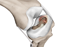
Meniscal Injuries
Meniscal tears are one of the most common injuries to the knee joint. It can occur at any age but are more common in athletes involved in contact sports. The meniscus has no direct blood supply and for that reason, when there is an injury to the meniscus, healing is difficult.
-
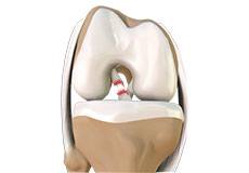
Articular Cartilage Injury
Articular or hyaline cartilage is the tissue lining the surface of the two bones in the knee joint. Cartilage helps the bones move smoothly against each other and can withstand the weight of the body during activities such as running and jumping.
-
Patellar Instability
Any damage to the supporting ligaments may cause the patella to slip out of the groove either partially (subluxation) or completely (dislocation). This misalignment can damage the underlying soft structures such as muscles and ligaments that hold the kneecap in place.
-
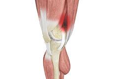
Knee Ligament Injuries
The knee is a hinge joint made up of two bones, the thighbone (femur) and shinbone (tibia). Ligaments are tough bands of tissue that connect one bone to another bone. The ligaments of the knee stabilize the knee joint.
-
Multiligament Knee Injuries
Injury to more than one knee ligament is called a multiligament knee injury and may occur during sports or other physical activities.
-
Hoffa's Fat Pad Syndrome
Hoffa’s fat pad syndrome also called fat pad impingement, infrapatellar fat pad syndrome, and Hoffa's disease, is a condition characterized by anterior knee pain, pain in the center, and front of your knees, due to inflammation of the Hoffa’s fat pad.
-
Iliotibial Band Syndrome
An iliotibial band is a tough group of fibers that runs from the iliac crest of the hip along the outside of the thigh, till the outer side of the shinbone, just below the knee joint. Its function is to coordinate with the thigh muscles and provide stability to the knee joint.
-
Jumper's Knee
Jumper’s knee, also known as patellar tendinitis, is inflammation of the patellar tendon that connects your kneecap (patella) to your shinbone. This tendon helps in the extension of the lower leg.